American Head & Neck Society

Advancing Education, Research, and Quality of Care for the Head and Neck oncology patient.

  • About
    • Mission Statement and Purpose
    • Divisions & Services of the Society
      • Education
        • Scientific Program/Resident Courses
        • Surgical Videos
        • Journal Club
        • Journals
        • Global Outreach
        • Awards
          • Margaret F. Butler Award
      • Diversity, Equity and Inclusion Division
      • Patient Care
        • Cancer Survivorship
          • Patient Education on Post-Treatment Care
          • Interviews with Cancer Survivors
        • Cancer Prevention
          • SLIDE DECK: HPV-Related Oropharyngeal Cancer
        • Guidelines/Position Statements
        • Find-A-Physician
      • Research
        • Grant Information
        • Clinical Trial
        • Tissue Banks
      • Administrative Division
        • AHNS Process for Evaluating Projects Requiring Funding
    • Leadership
    • History
      • Society Background
      • AHNS History Interviews
      • Past Presidents
      • In Memory
    • AHNS Newsletter
    • Professionalism & Ethics
    • AHNS Policies and Procedures (P&P) Manual
    • AHNS Foundation
    • AHNS Bylaws
    • AHNS Staff
    • AHNS News and Announcements
    • COVID-19 Bulletin Board
  • Heads Up!
  • Post a Job
  • Meetings
    • AHNS Virtual Education Series
      • TORS Webinar Series
    • AHNS Meetings Info
    • AHNS Call For Abstracts
    • Past Meetings
    • Related Meetings
  • For Patients
  • For Trainees
    • Fellowship Curriculum for FY2024-2025
      • For Current AHNS Fellows
      • Certificate of Completion Request
    • Accredited Fellowships
      • Directory of Fellowships
      • AHNS Fellowship Match
      • Fellowship Curriculum
      • Fellowship Graduates
      • For Program Directors
    • AHNS Surgical Videos
    • Fellows’ Virtual Tumor Boards
    • Cutaneous Cancer
  • Sections
    • Endocrine Surgery
    • Skull Base Surgery Section
    • Reconstructive Head & Neck Surgery
      • Head and Neck Reconstruction- Information for Patients
    • Mucosal Malignancy Section
      • Mucosal Malignancy Section Patient Information
    • Salivary Gland Section
      • Salivary Gland Section Patient Education Handout: Parotidectomy
    • Cutaneous Cancer
  • Find-A-Physician
  • Member Central
    • Join AHNS
    • Find-A-Physician
    • Mailing List Order
  • Login
    • Password Reset
  • Donate

Published on February 11, 2016 by AHNS Webmaster

2016 Prevention & Early Detection / Community Service Grants – Applications due February 29, 2016

The American Head and Neck Society’s Prevention and Early Detection Committee is pleased to open the application process for its 2016 Community Service grants. These grants are awarded in support of a project or community activities related to Oral Head and Neck Cancer Awareness (OHANCA) Program. Each grant, in the amount of $1,000.00, will be given to an individual, department, organization, or institution in support of a patient or community-oriented project held in conjunction with the annual Oral Head & Neck Cancer Awareness Week, taking place April 10-16, 2016.

A total of three grants are awarded – two AHNS Community Service Grants and a third international grant funded by the Head and Neck Cancer Alliance – the HNCA/AHNS International Outreach Cancer Prevention Grant.

Applicants should submit a letter, not to exceed 2 typed pages, containing a detailed description of the project, which should include:

  • The targeted population;
  • The methods to be used;
  • The expected outcome;
  • The expected impact on community health and/or on our knowledge and understanding of head and neck cancer prevention and early detection;
  • The estimated/actual cost, of the project;
  • Any other available funding.
  • Please also include a contact name, address, phone number and email.

Submit your application to:
Terri Lin-White, AHNS Administrative Associate
American Head and Neck Society
11300 W. Olympic Boulevard, Suite 600
Los Angeles, CA 90064
E-mail: [email protected]
Telephone: 310-437-0559 ext 172

Applications are due Monday night, February 29st at 11:59pm Eastern Time. Recipients will be announced in mid March.

Please note that indirect support and salary are not supported as part of this grant.

Recipients of this award, upon completion of their project, will be requested to submit a brief report describing how the funding was spent, the results of the project and an overall evaluation.

Published on January 21, 2016 by AHNS Webmaster

Neck Dissection

Authored by Nilesh Vasan, MD & Arjun Joshi, MD; edited by Miriam Lango, MD

AHNS Education Committee

What is a Neck Dissection and Why is it Performed?

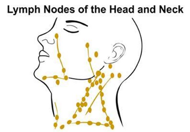
Neck dissection is usually performed to remove cancer that has spread to lymph nodes in the neck.lymph 1

Lymph nodes are small bean shaped glands scattered throughout the body that filter and process lymph fluid from other organs. The immune cells in the lymph nodes help the body fight infection. When cancer cells spread from another part of the body, they may get caught in a lymph node where they grow. An individual might feel a non-tender lump in the neck. The cancer in the lymph node is known as a metastasis. When someone undergoes surgery for cancer that has spread to lymph nodes, both the initial or primary cancer as well as the metastases must be removed.

Neck dissection refers to the removal of lymph nodes and surrounding tissue from the neck for the purpose of cancer treatment. The extent of tissue removal depends on many factors including, the stage of disease which reflects the extent of cancer as well as the type of cancer. The most common cancers removed from lymph nodes in the neck include head and neck squamous cell carcinomas, skin cancers including melanoma and thyroid cancers.. In general, the goal of neck dissection is to remove all the lymph nodes within a predefined anatomic area. Many of the lymph nodes removed during surgery will not prove to have cancer in them.

lymph 2Many patients wonder why so many non-cancerous lymph nodes must be removed; why can’t surgery be done to remove only the lymph nodes with cancer in them?

A cancer may shed any number of metastases that lodge in lymph nodes, grow and spread. There are over 150 lymph nodes on each side of the neck. During an operation, a surgeon will not be able to tell if a lymph node is clean, or if it has cancer that will later grow into a visible neck lump. The lymph nodes must be processed and tested; this takes time. For that reason, it is recommended that the lymph nodes in a predefined region are removed, not just lymph nodes that are obviously enlarged with cancer.

In addition, different cancers spread differently. Skin cancers first spread to lymph nodes in different parts of the neck than thyroid cancers or oral or larynx cancers. Squamous cell carcinomas that start in the lining or mucosa of the mouth, throat or larynx have a tendency to spread to lymph nodes early; cancer cells can often be detected in lymph nodes in the neck when examined under the microscope, even in the absence of visible or palpable neck lumps.

Lymph node metastasis reduces the survival of patients with squamous cell carcinoma by half. The survival rate is less than 5% in patients who previously underwent surgery and have a recurrent metastasis in the neck. Therefore, the control of cancer that has spread to the neck is one of the most important aspects in the successful management of these particular cancers. The neck dissection is a standardized procedure that was developed to ensure the complete removal of cancer that has spread to the lymph nodes of the neck.

Radical vs. Modified Neck Dissection

Radical Neck Dissection

  • This operation has been used for almost 100 years and describes the removal of lateral neck nodes and tissues to surgically remove cancer in the neck. Included in this tissue, which extends from the collarbone (clavicle) inferiorly to the jawbone (mandible) superiorly are dozens of lymph nodes. In addition to lymph nodes, this operation often includes remove the submandibular gland (a saliva-producing gland in the upper neck), the sternocleidomastoid muscle, the jugular vein and the spinal accessory nerve (to the trapezius muscle).
  • The radical neck dissection was designed to ensure complete cancer removal in individuals with very advanced cancers in the neck. The radical neck dissection is effective but produces characteristic cosmetic changes. Because so much tissue is removed, one side of the neck may appear flatter than the other. More importantly, sacrifice of the nerve to the trapezius muscle results in visible drooping of the shoulder and difficulty in raising the arm over head. Some individuals develop pain in the neck and collarbone. An intensive physical therapy program may diminish some of these problems, but some long-term disability can be anticipated.
  • Historically, radical neck dissections were the most commonly performed type of neck dissection performed by surgeons. This is no longer the case. Most patients are candidates for a less radical operation.

Modified Radical Neck Dissection

  • This term describes a variety of neck dissections that preserve structures that are usually sacrificed in the radical neck dissection such as the spinal accessory nerve, the internal jugular vein or sternocleidomastoid muscle. Further, selective neck dissections are neck dissections that, in addition to preserving these important structures, are used to remove specific groups of lymph nodes, rather than all the lymph nodes on the side of the neck, based on the probability that those lymph nodes harbor cancer.
  • While radical neck dissections produce the greatest changes in cosmetic appearance and shoulder function, selective neck dissections produce the least. In fact, selective neck dissections frequently produce no obvious cosmetic changes, yielding a nearly invisible scar. Nevertheless, strength and flexibility may be enhanced with adherence to neck and shoulder range of motion exercises after surgery. The best results can be expected with faithful adherence to an exercise program over the long term.

The Procedure

Neck dissections are done under general anesthesia through an incision that runs along a skin crease in the neck, extending vertically on the side of the neck. Incisions are usually designed to enhance the visualization and protection of important structures in the neck, and enable the safe removal of lymph nodes that harbor cancer.

Beneath the skin, underlying fat, and a thin layer of muscle (the platysma), the dissection proceeds to identify and remove the tissue containing the lymph nodes. If the sternocleidomastoid muscle is removed as part of the operation, there may be some flattening of the neck, but removal of this muscle rarely results in significant weakness.

What are the risks of neck dissection?

Neck dissections are subject to numerous potential operative complications that are common to all operative procedures, as well as complications specific to this procedure. Some of these are described below, but do not include all potential complications associated with neck dissection. The risk of specific complications may be best determined for an individual by the nature and extent of their cancer, prior treatment and other circumstances.

  • Bleeding-Patients may bleed after an operation. Bleeding under the skin after a neck dissection is rare. Sometimes an operative procedure to remove the blood is required. Rarely, a blood transfusion is also needed.
  • Infection can occur after any surgical procedure including neck dissection (uncommon)
  • Chyle leak, which results in fluid accumulation in the neck from disruption of the thoracic duct (this problem is more common after left sided neck dissections) (rare)
  • Wound healing problems requiring additional surgery (rare)

Several important nerves are found in the neck around the lymph nodes, and depending on the area of the neck to be operated, these nerves can be at risk for damage.   The primary nerves of concern are-

  • The marginal nerve, a small branch of the facial nerve which controls lower lip movement
  • The spinal accessory nerve which aids in shoulder mobility and raising the arm over head
  • The hypoglossal nerve, which controls movement of the tongue (uncommon)
  • The lingual nerve, which controls sensation on the side of the tongue (rare)
  • The vagus nerve which controls movement of one vocal cord (rare)

Additional potential long-term problems include:

  • Incision-Most incisions heal well, but some individuals develop scars.
  • Numbness of the skin along the incision as well as over the cheek, ear and neck can be anticipated which improves with time; some long term numbness can be anticipated
  • Neck stiffness or pain
  • Long term swelling in the neck or lymphedema
  • Shoulder weakness (uncommon)
  • Changes in speech and swallowing (rare)

Some problems are attributable to nerve injury; more commonly, scarring under the skin from surgery and radiation contributes to disability. Some problems may be avoided with early and faithful adherence to a shoulder range of motion exercise program, lymphedema or speech therapy rehabilitation programs.

Published on January 21, 2016 by AHNS Webmaster

Cancer of the Oropharynx: Risk Factors, Diagnosis, Treatment, and Outcomes

Authored by Charles Coffey, MD & Tamer Ghanem, MD PhD; edited by Ellie Maghami, MD FACS

AHNS Education Committee

Introduction

This document is intended to introduce a subtype of head and neck cancer known as oropharynx cancer, and the normal anatomy and physiology of the oropharynx will be reviewed. Factors that can lead to cancer formation in this area are defined. Information regarding diagnosis, treatment, and expected outcomes of this condition is provided.

Anatomy & Functional Considerations

The oropharynx is the middle compartment of the pharynx,  i.e. throat; it is the region of the throat between the nasopharynx (top compartment) and hypopharynx (bottom compartment). The oropharynx includes the tonsils, tongue base, soft palate, and pharyngeal walls. Several of these subsites are very common locations for the development of head and neck cancer, and a tumor present in any of these subsites can be broadly categorized as an oropharyngeal tumor. It is important to distinguish the oropharynx from the oral cavity (mouth), as tumors arising in these two sites may behave differently and require very different considerations regarding function and appropriate treatment.

The tonsils (also referred to as palatine tonsils) are collections of lymphoid tissue located on each side of the oropharynx which participate in the immune function of the aerodigestive tract. Although tonsils may be quite large during childhood, they generally regress with age, and many adults have very little visible tonsillar tissue remaining. Enlargement or asymmetry of the tonsils in an adult may simply be an anatomic variant, but may also be an indication of tumor presence. Removal of tonsils has not been found to compromise immune status.

The base of tongue (or tongue base) refers to the portion of the tongue which resides in the oropharynx (posterior 1/3 of the tongue). The base of tongue is functionally and anatomically distinct from the oral tongue, which is the portion of the tongue which resides in the oral cavity (anterior 2/3 of the tongue) and is most important for speech and language. The muscles of the base of tongue are more involved with swallowing than speech, and play a critical role in controlling the passage of food and liquids from the mouth into the throat. Base of tongue dysfunction resulting from tumor, loss of tissue due to surgery, or radiation-related effects may result in difficulty swallowing or aspiration (spillage of liquids into the larynx or voicebox). 

The soft palate is a muscular soft tissue sling which resides behind the hard palate, or roof of the mouth. The soft palate separates the nose and nasopharynx from the remainder of the pharynx and oral cavity during speech and swallowing. Inability to close the soft palate (velopharyngeal insufficiency) due to tumor, resection, or scar may result in hypernasal speech as well as reflux of liquids into the nose during swallowing.  The lateral and posterior walls of the oropharynx are comprised primarily of muscles which play a supporting role in the pharyngeal phase of swallowing.

Epidemiology

The oropharynx is one of the most common sites of head and neck cancer in the United States. It is estimated that 11,000-13,000 new cases of oropharyngeal cancer are diagnosed in the United States each year (Jemal; Siegel; CDC). The vast majority of these tumors are squamous cell carcinoma, a cancer which arises from squamous epithelial cells which line the upper aerodigestive tract.  Squamous cell carcinomas may also arise at numerous other sites including the skin, lungs, bladder, and cervix, but tumor behavior and treatment options vary greatly across different body sites.  Males are more than four times as likely as females to develop oropharyngeal squamous cell carcinoma (OPSCC), with an overall annual risk of 6.2 per 100,000 men compared to 1.4 cases per 100,000 women (Jemal, CDC). Incidence in the U.S. is highest among white and black men, and lower among Hispanics, Native Americans and Asians/ Pacific Islanders.

Risk Factors

Tobacco and Alcohol

Tobacco use has been strongly established as the primary risk factor in the majority of head and neck aerodigestive tract cancers (Sturgis 04, Sturgis 07). Lifetime risk of developing head and neck cancer is increased 10-fold in smokers, and the magnitude of risk increases up to 25-fold for the heaviest smokers. Alcohol use is an independent risk factor for development of head and neck cancer. Although the risk is higher with chronic heavy alcohol use, there is some evidence that light alcohol use may also increase risk of developing oropharyngeal cancer (Bagnardi). Combined use of both tobacco and alcohol further increase cancer risk (Masberg). Historically, up to 90% of head and neck squamous cell carcinoma including OPSCC has been attributed to tobacco use and alcohol abuse (Sturgis 04). Tobacco use in the United States has been steadily declining for the last five decades, with the percentage of active smokers decreasing from 43% in 1965 to below 20% in 2010 (Mariolis, Skinner). Per capita alcohol use has shown a more modest decline, from 2.7 gallons per year in the mid-1980s to 2.26 gallons per year in 2010 (LaVallee). These trends have generally been paralleled by decreasing rates of head and neck cancer incidence and mortality.

Human Papilloma Virus

Despite decreasing rates of tobacco and alcohol use, the rates of oropharyngeal cancer have trended steadily upward for the last decade. This is primarily due to the increase in cancers related to infection with the human papilloma virus (HPV). Human papilloma viruses are a large group of related viruses which are spread through vaginal, oral and anal sex. HPV is the most common sexually transmitted infection in the United States, affecting more than half of sexually active individuals at some point during their lives (NCI). Several strains of HPV are associated with increased risk of developing cervical, genital, and oropharyngeal cancers. Infection of epithelial cells by high-risk strains of HPV is associated with production of viral proteins which may eventually interfere with the cell’s normal ability to suppress tumor growth. The immune system successfully eliminates HPV infection in most patients, and only a small portion of patients infected with high-risk HPV strains will develop an HPV-related cancer.  It is estimated that approximately 7% of those aged 14 to 69 years in the U.S. have oral HPV infection at any one time, as detected by an oral rinse.  Approximately half (3.7%) of those infections are with high-risk HPV strains (Gillison 2012; Sanders). The time lag between an oral HPV infection and the development of HPV-related oropharyngeal cancer is estimated at between 15 and 30 years. As such, the rise in OPSCC seen since the 1990s in large part reflects changes in sexual practices in the 1960s and 1970s.

The risk profile for HPV-related OPSCC differs from most head and neck cancers. When compared to patients with non-HPV tumors, patients with HPV-positive OPSCC are more likely to be young, white, higher socioeconomic status, non-smokers, and non-drinkers. Sexual history is strongly associated with HPV-positive cancers. Significant increase in risk of OPSCC has been associated with lifetime number of sexual partners, any history of oral sex, earlier age at sexual debut, infrequent use of barrier devices during sex, lifetime history of sexually transmitted disease, and, among men, with a history of same-sex sexual contact (Heck; Gillison 2008).

An association between head and neck cancer and marijuana use has been demonstrated, but it remains uncertain whether marijuana use is an independent risk factor for OPSCC (Gillison 2008). Poor oral health, including periodontal disease and tooth loss, has been implicated in the etiology of oral and oropharyngeal carcinoma, but the associations have been modest and the evidence has generally been inconclusive (Divaris). High dietary intake of fruit and vegetables may be somewhat protective for oral and pharyngeal cancer, though the evidence for this has not been conclusive (Lucenteforte).

Diagnosis & Evaluation

Any patient with a persistent mass of the neck or throat or with symptoms suggesting oropharygneal cancer should be referred to an otolaryngologist (ENT) or head and neck surgeon for further evaluation. Symptoms of oropharyngeal cancer may include throat pain, difficulty swallowing, weight loss, ear ache, voice change, and blood-tinged saliva. Initial evaluation consists of a detailed medical history and comprehensive head and neck examination, generally including examination of the pharynx and larynx with a small flexible endoscope performed in an office setting. Any suspicious tumors of the oropharynx should be biopsied for histopathologic evaluation. In many instances it is possible to perform biopsy in clinic if a lesion is easily accessible either directly through the mouth or via flexible endoscopy. However, in many instances evaluation will require additional examination and biopsy under general anesthesia in an operating room.  Such procedures can generally be completed in less than thirty minutes and may be performed on an outpatient basis in most instances.  In certain circumstances, it is possible to obtain adequate tissue for diagnosis via a needle biopsy (fine needle aspiration, or FNA) if a lymph node within the neck has become involved with cancer (i.e. a nodal metastasis).  Needle biopsy is most often perfomed in clinic and does not require anesthesia, although it may not provide the same degree of staging information as examination under anesthesia. Testing of biopsy specimens for HPV status is recommended in all cases. HPV-positive status predicts better outcomes with standard therapies in general, but this information should not currently be used to tailor decisions regarding treatment with the exception of carefully monitored clinical trials..

All patients with a confirmed diagnosis of OPSCC should undergo evaluation by a multidisciplinary treatment team. Imaging should be obtained to evaluate the primary tumor, involvement of lymph nodes in the neck, and for evidence of metastatic cancer spread beyond the head and neck. Providers may choose to order either computed tomography (CT scan) or magnetic resonance imaging (MRI) of the neck to evaluate the pharynx and lymph nodes in the neck. This scan should be performed with IV contrast in nearly all cases, with the exception of patients with impaired kidney function or an allergy to contrast dye. CT scan of the chest is also indicated in most cases, to evaluate for the presence of metastatic cancer in the lungs or lymph nodes of the chest.  Positron emission tomography (PET scan) is also being used with increased frequency for pretreatment evaluation, particularly for patients with advanced-stage disease.

As part of the pretreatment evaluation, all patients who elect to pursue radiation therapy should undergo dental evaluation prior to treatment.  As a result of the effects of radiation on bone and surrounding tissue, there is a significantly increased risk of bone-related complications associated with dental procedures which are performed following radiation therapy. In the most severe instances, dental extraction or infection after radiation may result in death of bone tissue (osteoradionecrosis) requiring surgical removal of bone. To minimize the risk of these complications, tooth extractions prior to radiation may be recommended if there is evidence of dental decay or advanced periodontal disease. Pretreatment nutrition and speech and swallowing evaluations should be provided to all patients. Patients experiencing significant weight loss or swallowing difficulty as a result of OPSCC may benefit from surgical placement of a feeding tube into the stomach prior to treatment. However, many patients are able to maintain an oral diet throughout the duration of treatment, and feeding tube placement is not required for all patients. Whenever possible, patients undergoing treatment for OPSCC should continue to eat and drink by mouth during the course of treatment, as long as this is approved as safe by the treatment team. Exercising pharyngeal muscles during treatment can help maintain normal swallowing function both throughout and after cancer therapy.

In a small subset of patients squamous cell cancer is diagnosed only by needle aspiration biopsy of an abnormally enlarged lymph node. In these instances physical exam, imaging and intraoperative biopsies may fail to reveal an obvious cancer in the throat. These cases are referred to as “unknown primary” as the site of primary cancer development remains hidden. In these instances it is particularly recommended that the needle aspirate be tested for HPV. HPV-positive status suggests oropharynx as the most likely site for cancer origin and predicts improved outcomes.

Staging

TABLE 1&2

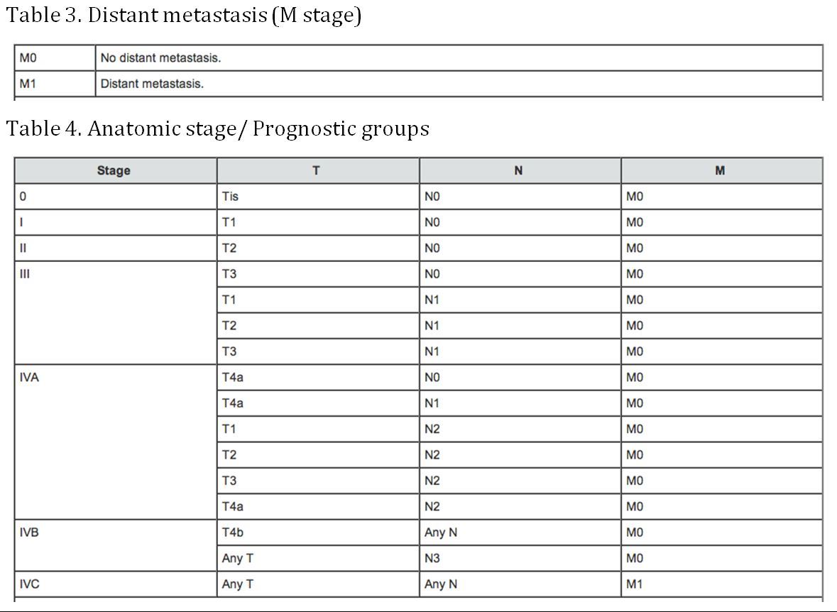
TABLE 3&4

(AJCC: Pharynx. In: Edge SB, Byrd DR, Compton CC, et al., eds.: AJCC Cancer Staging Manual. 7th ed. New York, NY: Springer, 2010, pp 41-56)

Pages: 1 2 3

Published on January 21, 2016 by AHNS Webmaster

Contemporary Understanding and Management of Oropharyngeal Carcinoma

Charles Coffey, MD; Tamer Ghanem, MD, David Goldenberg, MD FACS and Ellie Maghami, MD, American Head & Neck Society Education Committee

Introduction

This document is intended to introduce a subtype of head and neck cancer known as oropharynx cancer.  Normal anatomy and physiology of the oropharynx is reviewed. Factors that can lead to cancer formation in this area are defined. Information regarding diagnosis, treatment, and expected outcomes of this condition is provided.

Anatomy & Functional Considerations

The oropharynx is a critical functional component of the upper aerodigestive tract and is one of most common sites of origin of head and neck malignancy. For the purposes of functional and oncologic considerations it is important to distinguish the oropharynx from the oral cavity, which is a separate and distinct anatomic site. The oropharynx is comprised of the tonsils, base of tongue, soft palate, and pharyngeal walls. The soft palate is a muscular soft tissue sling which defines the superior extent of the oropharynx. It allows isolation of the nasopharynx from the remainder of the pharynx and oral cavity during speech and deglutition. Inability to close the soft palate (velopharyngeal insufficiency) due to tumor, resection, or fibrosis may result in hypernasal speech as well as nasopharyngeal reflux during swallowing. The palatine tonsils are collections of lymphoid tissue which reside on the lateral pharyngeal wall within a fossa formed by mucosal folds overlying the palatoglossus and palatopharyngeus muscles. Although the tonsils and associated lymphoid tissues of Waldeyer’s ring participate in humoral and cell-mediated immunity of the aerodigestive tract, removal of these tissues has not been demonstrated to compromise immune status (Böck ; Kaygusuz). The base of tongue comprises the ventral surface of the oropharynx, extending from the vallecula inferiorly to the circumvalate papillae anteriorly. Superficially lined by lymphoid tissue (lingual tonsils), the muscles of the base of tongue are critical in controlling passage of a food bolus during the pharyngeal phase of swallowing. Base of tongue dysfunction resulting from tumor, loss of tissue, denervation, or fibrosis may result in discoordination and delay in propulsion of food bolus, or premature spillage of liquids from the oral cavity into the pharynx and larynx. The later may result in aspiration.  The lateral and posterior walls of the oropharynx are comprised primarily of the superior and middle constrictor muscles, which play a supporting role in the pharyngeal phase of swallowing.

Epidemiology

The oropharynx is one of the most common sites of head and neck cancer in the United States, currently trailing only the oral cavity in annual incidence among head and neck cancers (Siegel). The vast majority of these tumors are squamous cell carcinoma, an epithelial malignancy which may affect any tissue site lined with squamous epithelium. The incidence of oropharyngeal squamous cell carcinoma (OPSCC) in the U.S. is estimated at between 11,000-13,000 new cases per year (Jemal; Siegel; CDC). Males are more frequently affected, with an overall annual risk of 6.2 per 100,000 men compared to 1.4 cases per 100,000 women (Jemal, CDC).

The incidence of OPSCC has risen sharply over the last several decades, due primarily to increased rates of tumors associated with the human papilloma virus (HPV). Although the incidence of HPV-negative tumors in the U.S. declined by 50% from 1988-2004 (paralleling the decrease in smoking and smoking-related malignancies), the incidence of HPV-positive tumors increased by 225% during the same time frame (Chaturvedi). The proportion of oropharyngeal cancers in the U.S. which were HPV-positive increased from 16% in the period from 1984-89 to 72% in the period from 2000-04 (Chaturvedi). Similar trends have been seen elsewhere. Rates of OPSCC in Australia increased by 43% between 1982 and 2008 (Ariyawardana), and the incidence of tosillar cancer in Swedish men increased at a rate of 2.6% per year from 1960-2003 (Hammarstedt). The rate of HPV-positive tumors in Sweden nearly doubled each decade from 1970-2007 (Näsman). After steadily declining during the previous 3 decades, the age-adjusted mortality of oropharyngeal carcinoma in the U.S. has trended gradually upward since 2000 (0.38/100,000 deaths in 2000; 0.46/100,000 in 2010) (Jemal). This overall increase in mortality is a reflection of increased incidence of these tumors rather than a decrease in survival rates.

Historically, oropharyngeal carcinoma was primarily a disease of older individuals, with highest incidence over the age of 70 (Hammarsted). However, these demographics have shifted in the era of HPV-associated OPSCC. The last two decades have seen a steady increase in the rates of HPV-positive OPSCC among younger individuals, most notably white males between the ages of 50 and 70 (Chaturvedi, CDC).

Risk Factors

Oropharyngeal carcinoma rates are 4-fold higher in men than women across ethnicities. Incidence in the U.S. is highest among white and black men (age-adjusted incidence of 8.5/100,000 and 7.9/100,000, respectively), and lower among Hispanics (5.7/100,000 men), Native Americans (4.7/100,000 men), and Asians/ Pacific Islanders (2.2/100,000 men) (Siegel, Jemal).

Tobacco use has been strongly established as the primary carcinogenic factor in the majority of head and neck aerodigestive tract cancers (Sturgis 04, Sturgis 07). Lifetime risk of developing head and neck cancer is increased 10-fold in smokers, and the magnitude of risk increases in a dose-dependent fashion, up to 25-fold for the heaviest smokers. Alcohol use is an independent risk factor for development of head and neck cancer. The most significant risk is associated with chronic heavy alcohol use, though there is some evidence that light alcohol use may also increase risk of developing oropharyngeal cancer (Bagnardi). Though tobacco and alcohol themselves are independent risk factors, the risks associated with combined tobacco and alcohol use are synergistic (Masberg). Historically, up to 90% of head and neck squamous cell carcinoma including OPSCC has been attributed to tobacco use and alcohol abuse (Sturgis 04).

Tobacco use in the United States has been steadily declining for the last five decades. The percentage of active smokers in the U.S. decreased from 43% in 1965 to 21% in 2005 (Mariolis), and that number dropped below 20% in 2010 (Skinner). Per capita alcohol use has shown a more modest decline, from 2.7 gallons per year in the mid-1980s to 2.26 gallons per year in 2010 (LaVallee). These trends have generally been paralleled by decreasing rates in head and neck cancer incidence and mortality. However, as outlined in the previous section, rates of oropharyngeal cancer have trended steadily upward for the last decade, primarily due to the increase in HPV-related tumors. Since the first description of the association between HPV and head and neck cancer in 1983, the role of HPV in the development of squamous cell carcinoma has become well established (Syrjänen; Gillison 2004; Li). The primary molecular mechanism of HPV-related carcinogenesis in OPSCC is deactivation of tumor suppressor genes p53 and Rb by the viral oncogenes E6 and E7, respectively (Li; Rothenberg). A number of high-risk viral subtypes have been identified, most notably HPV-16, which is present in up to 95% of HPV-related oropharyngeal tumors (Gillison 2008).

The risk profile for HPV-related OPSCC differs from most head and neck cancers, supporting clinical and molecular evidence that this is a distinct pathologic entity (Gillison 2004). When compared to HPV-negative head and neck cancer patients, patients with HPV-positive OPSCC are more likely to be young, white, higher socioeconomic status, non-smokers, and non-drinkers. Sexual history is strongly associated with HPV-positive cancers. Significant increase in risk of OPSCC has been associated with lifetime number of sexual partners including oral sex partners, any history of oral sex, earlier age at sexual debut, infrequent use of barrier devices during sex, lifetime history of sexually transmitted disease, and, among men, with a history of same-sex sexual contact (Heck; Gillison 2008). The sexual risk factors associated with oral HPV infection mirror those seen with HPV-related OPSCC. Current prevalence of oral HPV infection in the U.S. among individuals aged 14 to 69 years is approximately 7%, while incidence of oncogenic (high risk) subtypes is 3% (Gillison 2012; Sanders). The time lag between an oral HPV infection and the development of HPV-related oropharyngeal cancer is estimated at between 15 and 30 years. As such, the rise in OPSCC seen since the 1990s may reflect changes in sexual practices in the 1960s and 1970s.

An association between head and neck cancer and marijuana use has been demonstrated by Gillison et al. (Gillison 2008). This association was limited to HPV-positive tumors, and although it is not clear that marijuana use is an independent risk factor for OPSCC, it is plausible that cannabinoids may promote progression of HPV-mediated carcinogenesis via immunologic mechanisms.

Poor oral health, including periodontal disease and tooth loss, has been implicated in the etiology of oral and oropharyngeal carcinoma, but the associations have been modest and the evidence has generally been inconclusive (Divaris). Recent evidence has demonstrated increased risk of oral HPV infection related to poor oral health  and irrespective of smoking or oral sex history (Bui). This suggests that poor oral health may be an indirect risk factor for OPSCC, though this has not been borne out in other studies (Gillison 2008).

High dietary intake of fruit and vegetables may be somewhat protective for oral and pharyngeal cancer, though the evidence for this has not been conclusive, and at least part of the protective effect may be explained by confounding effects of tobacco and alcohol exposure (Lucenteforte).

Diagnosis & Evaluation

All patients with findings suspicious for oropharyngeal cancer should be referred to a head and neck surgeon or otolaryngologist for further evaluation. Initial evaluation consists of a detailed history and comprehensive head and neck examination, generally including flexible fiberoptic nasopharyngoscopy and laryngoscopy performed in an office setting. Any suspicious tumors of the oropharynx should be biopsied for histopathologic evaluation. In many instances it is possible to obtain an adequate tissue sample in clinic if a lesion is easily accessible either directly through the mouth or via flexible endoscopic biopsy. However, examination and biopsy under anesthesia may be required if achieving exposure for biopsy is more difficult; if medical considerations such as anticoagulation status preclude safe biopsy in an office setting; if airway intervention such as tracheostomy is required due to obstructive tumor; or if the clinic exam cannot provide sufficient evaluation of the primary tumor for accurate staging.  In select circumstances, cytopathologic evaluation via fine needle aspiration of a metastatic neck node may prove adequate tissue to establish diagnosis, provided that the tumor can be accurately clinicaly staged without exam under anesthesia. Testing of specimens for HPV status is recommended in all cases, either through immunohistochemistry for p16 expression or in situ hybridization for detection of HPV DNA. Although HPV-status should not be used in treatment planning outside of a clinical trial setting, information regarding HPV-status is currently the single greatest prognostic factor in evaluation of OPSCC.

Once a diagnosis of OPSCC is confirmed, all patients should undergo multidisciplinary evaluation. Imaging should be obtained to evaluate the primary tumor, nodal basin, and chest. Computed tomography (CT) and magnetic resonance imaging (MRI) of the neck both allow appropriate diagnostic imaging of the pharynx and cervical nodal basins, and either modality may be selected based upon the preference of providers and patients. Either iodinated contrast (CT) or gadolinium (MRI) should be administered unless medically contraindicated. A diagnostic imaging study of the neck should extend from the skull base to the thoracic inlet, and will thus provide evaluation of both the pharynx and nodes with a single study. There is no routine role for imaging of the head or brain in the evaluation of oropharyngeal cancer. Imaging of the chest generally consists of diagnostic-quality chest CT, although PET/CT may be considered in evaluation of advanced-stage disease (Stage III-IV) due to increased risk of distant metastasis.

Patients considered for radiation therapy should undergo dental evaluation prior to treatment, due to the increased risk of mandibular osteoradionecrosis associated with dental procedures (extraction, e.g.) when performed following radiation. If there is evidence of dental decay or advanced periodontal disease, prophylactic extraction prior to undergoing radiation is frequently recommended. Pretreatment nutrition and speech and swallowing evaluations should be provided to all patients. Patients experiencing significant weight loss or swallowing difficulty may benefit from feeding tube placement prior to treatment, although many patients are able to maintain an oral diet throughout the duration of treatment with either surgery-based or radiation-based approaches.

Staging

TABLE 1&2

TABLE 3&4

(AJCC: Pharynx. In: Edge SB, Byrd DR, Compton CC, et al., eds.: AJCC Cancer Staging Manual. 7th ed. New York, NY: Springer, 2010, pp 41-56)

Treatment

Treatment paradigms

Current NCCN guidelines for management of OPSCC  may be broadly divided based upon the distinction between early-stage (I-II) and advanced-stage (III-IV) disease.  Early-stage disease may generally be treated with single-modality therapy, consisting of either surgery or definitive radiation. Either modality should include management of both the primary tumor site within the oropharynx and also the at-risk cervical nodal basin. Treatment of the cervical nodes is required even for early-stage (clinically N0) disease due to the elevated risk for occult nodal metastasis associated with tumors of the oropharynx (Byers; Shah). If surgery is employed for early-stage disease, the addition of pathologic staging information helps guide whether additional treatments are warranted. For example, adjuvant radiation therapy (also referred to as postoperative radiation therapy, or PORT) may be recommended for select T1 and T2 disease when histopathologic evaluation demonstrates intermediate-risk adverse features such as perineural invasion, multiple metastatic lymph nodes, or close surgical margins.  Adjuvant chemotherapy may be employed when high-risk adverse features are identified, included the presence of extracapsular nodal disease or positive surgical margins(Cooper, Pajak et al. 2004) .

Advanced-stage tumors of the oropharynx require multimodality therapy. This may include primary surgery followed by adjuvant radiotherapy, or concurrent chemoradiation. Alternatively, induction chemotherapy may be employed prior to definitive radiotherapy or chemoradiation in an attempt to minimize risk of distant metastasis in patients considered to be at high-risk due to extensive primary tumor or high-volume nodal disease  (Posner and Vermorken 2008, Vermorken 2010).   In either setting, the primary effect of cytotoxic chemotherapy is to serve as a radiosensitizer. If residual tumor remains either at the primary site or within the neck following completion of definitive chemoradiation, surgery may be employed (Zafereo, Hanasono et al. 2009, Zafereo 2014) in an attempt to eradicate residual or recurrent disease. However, long-term outcomes for salvage surgery following failure of primary chemoradiation for OPSCC are often poor .

The current standard-of-care for administration of radiation to the orpharynx is intensity modulated radiation treatment (IMRT).  This technique utilizes sophisticated imaging to construct a three-dimensional map defining the treatment dose to the tumor and surrounding tissues. The advantage of IMRT over older techniques is the ability to optimize radiation delivery to the tumor site while minimizing dose to surrounding critical structures such as the spine, carotid arteries, and brain, and increasingly to spare portions of functionally critical structures such as the salivary glands and superior constrictor muscles .  The efficacy of IMRT over conventional radiation for treatment of oropharyngeal cancer has yet to be well established.  There are several dose-dependent side effects of head & neck radiation. Patients commonly complain of xerostomia, loss of taste, dysphagia (in some cases require feeding tube placement), fibrosis of neck skin and muscles, and oral and oropharyngeal mucositis. Late side effects include increased risk of carotid atherosclerotic disease,  esophageal stricture, osteoradionecrosis of the mandible, and the possibility of a radiation induced malignancy.

Surgical approaches

Surgery of oropharyngeal tumors can be performed through various approaches, to obtain access to the oropharynx dictated by the location and size of the tumor as well as functional and aesthetic concerns.  Traditional “open” approaches to the oropharynx include a transmandibular approach via lip-splitting incision and mandibulotomy or transcervical approaches involving neck dissection and incisions into the anterior or lateral pharynx (Coffey; Mehta).  These approaches offer excellent surgical exposure of the tonsillar fossa, soft palate, and base of tongue, though they are often accompanied by significant functional morbidity due to the extensive dissection required.  The majority of these patients require short-term tracheostomy and/ or feeding tube placement, and soft tissue reconstruction with a vascularized flap is often require to close the surgical defect and reduce the risk of fistula formation. Reconstruction frequently consists of regional flaps such as the pectoralis major flap or various microvascular flaps, each of which is accompanied by additional morbidities.  Additional disadvantages of these open approaches includes prolonged operative times, significant postoperative edema, external scarring, and the potential for mandibular complications including malunion, nonunion or bite abnormality.  Given the increasing use and proven efficacy of radiation therapy, most centers in the United States currently reserve open surgical approaches for salvage cases where additional radiation is not possible or too risky, and where less invasive surgical approaches could compromise oncologic outcomes.

The last several decades have seen the advent of transoral surgical approaches for management of OPSCC.  Although very early stage lesions of the tonsil or base of tongue may be resectable with very basic equipment and minimal technical difficulty, the challenging anatomy and difficult exposure of the oropharynx often preclude such straightforward approaches.  However, recent technical advances have allowed surgeons to resect even relatively large tumors of the oropharynx via strictly transoral approaches, most notably including  transoral laser microsurgery (TLM), or transoral robotic surgery (TORS).  Transoral laser microsurgery was popularized by Steiner (representative citations), and combines conventional suspension laryngoscopy techniques with microscopic magnification and CO2 laser to remove tumors of the oropharynx and larynx.  The advantages of TLM include improved access and visualization in difficult areas such as suprglottis and glottis, and relative availability of the required instrumentation.  Disadvantages of TLM include a steep learning curve and the limitations of line-of-sight laser application.

In December of 2010, TORS was approved by the FDA for management of T1 and T2 oropharyngeal and supraglottic cancers, and this approach is gaining increasing popularity in the U.S. and abroad.  TORS capitalizes on the abilities conferred by the Da Vinci Robot (Intuitive Surgical), including three-dimensional visualization of the target anatomy and the ability to work precisely in a confined space such as the oropharynx.  The TORS surgeon sits on a console allowing control the movement of robotic arms, which are introduced to the surgical site through the mouth with the aid of a retractor to obtain exposure of the oropharynx.    The primary advantages of this approach include more rapid learning curve than TLM, ability to resect tumor en bloc instead of piecemeal, excellent three-dimensional magnified view of the relevant anatomy, and precise articulated instrumentation.  The disadvantages include limited availability due to need for expensive surgical equipment and specialty training of the surgical and OR staff.  Numerous studies on the oncological efficacy of this approach have been published, demonstrating excellent oncologic control equivalent to or better than standard surgical and non-surgical techniques (References- consider White 2013; de Almeida 2014; others)). 

The general advantages of transoral surgical approaches include decreased morbidity compared with more extensive open dissections, avoidance of tracheostomy, decreased blood loss, and shorter surgical times.  (Include data/ citations regarding specific functional outcomes; consider More 2013). Decreased hospital stays are often possible as well (White 2013).  However, due to anatomic and technical constraints not all tumors are amenable to transoral resection, and these techniques also require considerable experience on the part of the surgeon, as well as instrumentation which may not be available at many centers.

Follow up & Outcomes

Nonsurgical Treatment

One of the difficulties in comparing outcomes between surgical and non-surgical treatments for oropharyngeal cancer is lack of prospective randomized control trials to assess the efficacy and functional outcomes of the different available treatments.  This is an area currently being investigated through RTOG and ECOG trials.  However, the trend in the past 10-15 years has been use of concomitant chemoradiation therapy. There are several studies that have evaluated the response of combined radiation and chemotherapy on oropharyngeal cancer.  In a large recently published single-institution study, 1046 patients with oropharygneal cancer were treated at a single institution with non-surgical treatment (radiation alone, concomitant chemoradiation, or induction chemo followed by chemoradiation) 5-year year overall survival, local control, regional  control rates were 78%, 77%, and 87%, respectively (Garden).  An older, yet highly cited study, performed a meta-analysis of 51 published studies on oropharyngeal cancer from 1970-2000 (Parsons).  They found that the rates of cancer control between surgery followed by radiation therapy versus radiation therapy alone were similar:  local control 79% versus 76%, loco-regional control 60% versus 69%, 57% versus 59% 5-year  disease specific survival , respectively. While the cancer control was not significantly different between the cohorts, the rates of severe complications were much higher in the surgery group versus the primary radiation group, 23% vs. 6%.  Similarly the rates of fatal complications were higher in the surgery group versus the radiation group 3.2% versus 0.8%, respectively. The primary drawback of this study is the inclusion of older literature, which favored open surgical approaches with and without reconstructive techniques, and pre-modern radiation techniques such as intensity modulated radiotherapy therapy (IMRT).  Furthermore, newer literature supports adding chemotherapy to radiation to improve cancer control rates (Blanchard; Pignon 2007; Pignon 2009).  Unfortunately, the addition of chemotherapy to radiation therapy increases the toxicity profile of the treatment regimen, and can severely affect the patient’s quality of life especially in the areas of deglutition and xerostomia.

Surgical Treatment

Using open surgical approaches for base of tongue cancer ablation followed by adjuvant treatment, results are variable depending on T-stage of the disease.  In a study from Pittsburgh with 87 patients with base of tongue cancer, overall and disease specific survival at 5-years were 49% and 56%, respectively (Gourin and Johnson 2001).  For 5-year disease specific survival T1 was 88%,  T2 was 64%, T3 was 58%, and T4 was 30%.  Utilizing transoral surgical approaches, whether TLM or TORS, excellent tumor control can be achieved as reported by multiple investigators.   White and colleagues reported an 86.5% recurrence free survival at two years follow up in a cohort of 89 patients (T1, T2 n=79; T3, T4=18) (White, Moore et al. 2010).  In a separate group of 47 patients, treated TORS by Weinstein, disease specific survival was 90% at 2 years.  In this cohort, 38% avoided additional chemotherapy as a result of undergoing surgical treatment, and 11% of patients did not require adjuvant radiotherapy.  Also, one of out the 47 patients require gastrostomy tube placement at one year.  Similarly, the data in the TLM literature is very robust.  In a recent study by Patel and colleagues from Mayo Clinic, the locoregional control , recurrence free and overall survival rates at 3 years in a cohort of 80 patients (T1 52.5%, T2 38.8%, and T3 8.8%) were 98.6%, 91.1%,  and 93.7%, respectively.

References

Böck A, Popp W, Herkner KR.  Tonsillectomy and the immune system: a long-term follow up comparison between tonsillectomized and non-tonsillectomized children. Eur Arch Otorhinolaryngol. 1994;251(7):423-7.

Kaygusuz I, Alpay HC, Gödekmerdan A, et al. Evaluation of long-term impacts of tonsillectomy on immune functions of children: a follow-up study. Int J Pediatr Otorhinolaryngol. 2009 Mar;73(3):445-9.

Centers for Disease Control and Prevention (CDC). Human papilloma virus- associated cancers- United States, 2004-2008. MMWR Morb Mortal Wkly Rep 2012; 61:258-61

Siegel R, Ward E, Brawley O, et al. Cancer statistics, 2011: The impact of eliminating socioeconomic and racial disparities on premature cancer deaths. CA Cancer J Clin 2011; 61:212-36

Jemal A, Simard EP, Dorell C,  et al. Annual Report to the Nation on the Status of Cancer, 1975-2009, featuring the burden and trends in human papillomavirus (HPV)-associated cancers and HPV vaccination coverage levels. J Natl Cancer Inst. 2013 Feb 6;105(3):175-201

Syrjänen K, Syrjänen S, Lamberg M et al. Morphological and immunohistochemical evidence suggesting human papillomavirus (HPV) involvement in oral squamous cell carcinogenesis. Int J Oral Surg 1983; 12: 418–424

Näsman A, Attner P, Hammarstedt L, et al. Incidence of human papillomavirus (HPV) positive tonsillar carcinoma in Stockholm, Sweden: an epidemic of viral-induced carcinoma? Int J Cancer 2009 Jul 15;125(2):362-6

Hammarstedt L, Dahlstrand H, Lindquist D, Onelov L, Ryott M, Luo J, Dalianis T, Ye W, Munck-Wikland E. The incidence of tonsillar cancer in Sweden is increasing. Acta Otolaryngol 2007;127:988–92.

Ariyawardana A, Johnson NW. Trends of lip, oral cavity and oropharyngeal cancers in Australia 1982–2008: overall good news but with rising rates in the oropharynx. BMC Cancer 2013; 13:333

Chaturvedi AK, Engels EA, Pfeiffer RM, et al. Human papillomavirus and rising oropharyngeal cancer incidence in the United States. J Clin Oncol. 2011 Nov 10;29(32):4294-301

Sturgis EM, Cincirpini PM. Trends in head and neck cancer incidence in relation to smoking prevalence; an emerging epidemic of human papillomavirus-associated cancers? Cancer 2007; 110:1429-35

Li G, Sturgis EM. The role of human papillomavirus in squamous carcinoma of the head and neck. Curr Oncol Rep 2006; 8:130–139

Rothenberg SM, Ellisen LW. The molecular pathogenesis of head and neck squamous cell carcinoma. Journal of Clinical Investigation 2012; 122:1951-57

Sturgis EM, Wei Q, Spitz MR. Descriptive epidemiology and risk factors for head and neck cancer. Semin Oncol 2004; 31:726-33.

Blot WJ, McLaughlin JK, Winn DM, et al. Smoking and drinking in relation to oral and pharyngeal cancer. Cancer Res 1988; 48:3282-7.

Carvalho AL, Nishimoto IN, Califano JA, Kowalski LP. Trends in incidence and prognosis for head and neck cancer in the United States: a site-specific analysis of the SEER database. Int J Cancer 2005;114:806–808.

Schlecht NF, Franco EL, Pintos J, Kowalski LP. Effect of smoking cessation and tobacco type on the risk of cancers of the upper aero-digestive tract in Brazil. Epidemiology 1999; 10:412–418.

Mashberg A, Boffetta P, Winkelman R et al. Tobacco smoking, alcohol drinking, and cancer of the oral cavity and oropharynx among US veterans. Cancer 1993; 72: 1369–1375.

Bagnardi V, Rota M, Botteri E, et al. Light alcohol drinking and cancer: a meta-analysis. Annals of Oncology 2013; 24: 301–308

Mariolis P, Rock VJ, Asman K, et al. Tobacco use among adults- United States, 2005. MMWR Morb Mortal Wkly Rep 2006; 55:1145–1148

Gillison ML. Human papillomavirus-associated head and neck cancer is a distinct epidemiologic, clinical, and molecular entity. Semin Oncol 2004; 31:744–754

Gillison ML, D’Souza G, Westra W, et al. Distinct risk factor profiles for human papillomavirus type 16-positive and human papillomavirus type 16-negative head and neck cancers. J Natl Cancer Inst. 2008;100(6):407-20

Gillison ML, Broutian T, Pickard RK, et al. Prevalence of oral HPV infection in the United States, 2009-2010. JAMA 2012;307:693-703

Sanders AE, Slade GD, Patton LL. National prevalence of oral HPV infection and related risk factors in the U.S. adult population. Oral Dis 2012; 18:430-41

de Sanjosé S, Diaz M, Castellsagué X, et al. Worldwide prevalence and genotype distribution of cervical human papillomavirus DNA in women with normal cytology: a meta-analysis. Lancet Infect Dis 2007; 7:453-9

Heck JE, Berthiller J, Vaccarella S, et al. Sexual behaviours and the risk of head and neck cancers: a pooled analysis in the International Head and Neck Cancer Epidemiology (INHANCE) consortium. Int J Epidemiol. 2010; 39(1):166-81

Settle K, Posner MR, Schumaker LM. Racial survival disparity in head and neck cancer results from low prevalence of human papillomavirus infection in black oropharyngeal cancer patients. Cancer Prev Res 2009; 2:776-81

Skinner HD, Holsinger FC, Beadle BM. Oropharynx cancer. Curr Probl Cancer 2012; 36:334-415

LaValee RA, Yi H. Surveillance report #95: Apparent per capita alcohol consumption: National, state, and regional trends, 1977-2010. In: Alcoholism NIoAAa Arlington, TX: National Institute on Alcohol Abuse and Alcoholism, Division of Epidemiology and Prevention Research, 2012.

Divaris K, Olshan AF, Smith J, et al. Oral health and risk for head and neck squamous cell carcinoma: the Carolina Head and Neck Cancer Study. Cancer Causes Control 2010; 21:567-75

Bui TC, Markham CM, Ross MW, Mullen PD. Examining the association between oral health and oral HPV infection. Cancer Prev Res; 6(9) September 2013 (online ahead of print)

Lucenteforte E, Garavello W, Bosetti C, et al. Dietary factors and oral and pharyngeal cancer risk. Oral Oncol 2009;45:461-7

Byers RM, Wolf PF, Ballantyne AJ. Rationale for elective modified neck dissection. Head & Neck Surgery 1988; 10:160-7

Shah JP. Patterns of cervical lymph node metastasis from squamous carcinomas of the upper aerodigestive tract. The American Journal of Surgery 1990; 160:405-9

Cooper JS, Pajak TF, Forastiere AA, et al. Postoperative concurrent radiotherapy and chemotherapy for high-risk squamous-cell carcinoma of the head and neck. N Engl J Med 2004; 350:1937-44

Vermorken JB, Remenar E, van Herpen C, et al. Cisplatin, fluorouracil, and docetaxel in unresectable head and neck cancer. N Engl J Med 2007; 357:1695-704

Posner MR, Hershock Dm, Blajman CR, et al. Cisplatin and fluorouracil alone or with docetaxel in head and neck cancer. N Engl J Med 2007; 357:1705-1715

Haddad R, O’Neill A, Rabinowits G, et al. Induction chemotherapy followed by concurrent chemoradiotherapy (sequential chemoradiotherapy) versus concurrent chemoradiotherapy alone in locally advanced head and neck cancer (PARADIGM): a randomised phase 3 trial. Lancet Oncology 2013; 14:257-64

Zafereo ME, Hanasono MM, Rosenthal DI, et al. The Role of Salvage Surgery in Patients with Recurrent Squamous Cell Carcinoma of the Oropharynx. Cancer 2009, 115:5723-33

Coffey CS, Day TA. (2013) Cancer of the oral cavity: surgical technique. In Head and Neck Cancer: A Multidisciplinary Approach, 4th edition. Eds. Harrison LB, Sessions RB, Kies MS. Philadephia, PA: Lippincott, Williams, and Wilkins

Mehta V, Ferris RL. (2013) Cancer of the oropharynx: surgical technique. In Head and Neck Cancer: A Multidisciplinary Approach, 4th edition. Eds. Harrison LB, Sessions RB, Kies MS. Philadephia, PA: Lippincott, Williams, and Wilkins

Garden AS, Kies MS, Morrison WH, et al. Outcomes and patterns of care of patients with locally advanced oropharyngeal carcinoma treated in the early 21st century. Radiat Oncol 2013; 8:21. 

Parsons JT, Mendenhall WM, Stringer SP, et al. Squamous cell carcinoma of the oropharynx: surgery, radiation therapy, or both. Cancer 2002; 94:2967-2980.

Blanchard P, Baujat B, Holostenco Vet al. Meta-analysis of chemotherapy in head and neck cancer (MACH-NC): a comprehensive analysis by tumour site. Radiother Oncol 2011; 100:33-40.

Pignon JP, le Maitre A, Bourhis J. Meta-Analyses of Chemotherapy in Head and Neck Cancer (MACH-NC): an update. Int J Radiat Oncol Biol Phys 2007; 69:S112-114.

Pignon JP, le Maitre A, Maillard E, Bourhis J. Meta-analysis of chemotherapy in head and neck cancer (MACH-NC): an update on 93 randomised trials and 17,346 patients. Radiother Oncol 2009; 92:4-14.

Gourin CG, Johnson JT. Surgical treatment of squamous cell carcinoma of the base of tongue. Head Neck 2001; 23:653-660.

White HN, Moore EJ, Rosenthal ELet al. Transoral robotic-assisted surgery for head and neck squamous cell carcinoma: one- and 2-year survival analysis. Arch Otolaryngol Head Neck Surg 2010; 136:1248-1252.

Posner, M. and J. B. Vermorken (2008). “Induction therapy in the modern era of combined-modality therapy for locally advanced head and neck cancer.” Semin Oncol 35(3): 221-228.

Vermorken, J. B. (2010). “A new look at induction chemotherapy in locally advanced head and neck cancer.” Oncologist 15 Suppl 3: 1-2.

Zafereo, M. (2014). “Surgical salvage of recurrent cancer of the head and neck.” Curr Oncol Rep 16(5): 386.

Published on August 25, 2015 by AHNS Webmaster

Hands-on Transoral Laser Microsurgery Course – October 10 & 11, 2015

Presented by Memorial Sloan Kettering Cancer Center.

Hands-on Transoral Laser Microsurgery Course


For more information click here »

  • 1
  • 2
  • 3
  • 4
  • Next Page »

News and Announcements

  • AHNS YMCP Episode 11 – Melina Windon, MD, Janice Farlow, MD, Eric Gantwerker, MD May 8, 2025
  • Artificial Intelligence in Management of H&N Cancer: New Horizons Presented April 28, 2025
  • A conversation with Dr. Uppaluri: Neoadjuvant Therapy for Advanced H&N Mucosal April 25, 2025
  • The End of April Head and Neck Cancer Awareness Month Approaches! April 23, 2025
  • April is National CBD Awareness Month! April 21, 2025

AHNS Meetings and Events

AHNS 2025 Annual Meeting
Held during the Combined Otolaryngology Spring Meetings (COSM)

May 14-18, 2025
Hyatt Regency New Orleans
New Orleans, Louisiana

learn more...

Contact Us

AHNS, 11300 W. Olympic Blvd, Suite 600
Los Angeles, CA 90064
ph: (310) 437-0559 / fx: (310) 437-0585
[email protected]

Welcome to the AHNS Virtual Repository of Scientific Research

Sections

  • Virtual Repository of Scientific Resources
  • Cell Lines Browse and Search
  • Omics Browse and Search
  • Patient Derived Xenografts Browse and Search
  • Plasmids Browse and Search
  • Pre-Clinical Models Browse and Search
  • Transferable Expertise Browse and Search
  • Transgenic Mice Browse and Search

© 2002–2025 American Head and Neck Society · Privacy and Return Policy
· Managed by BSC Management, Inc